
Patient and presentation
A 26-year-old patient presented with a lower posterior tooth affected by caries for several years and requested restoration.
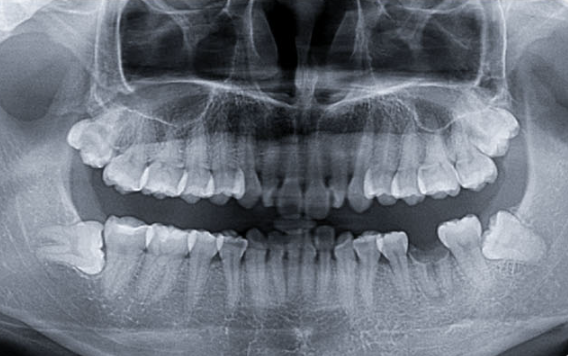
A residual crown at site 36 was covered by soft-tissue overgrowth. Imaging showed a widened periodontal space and a small periapical radiolucency consistent with suspected granulomatous pathology.
Extraction and planning
A minimally invasive extraction was completed, followed by delayed implant planning.
Preoperative CT showed incomplete socket remodeling, approximately 15.1 mm to the mandibular canal and 5.9 mm lingual bone width. A BL.RC4110 implant (Ø4.1 × 10 mm) was planned in the septal bone.
Implant placement and integration
The implant was placed with favorable postoperative three-dimensional position. At follow-up, the peri-implant tissue was healthy and the keratinized gingiva was normal.
After removal of the healing abutment, ISQ readings in the mesiodistal and buccolingual directions were above 70, supporting progression to restoration.
Restoration and review
A closed-tray impression was made and the definitive restoration was inserted with occlusal adjustment.
Nine months later, the gingival mucosa showed no reported abnormality and periapical imaging showed good peri-implant bone condition. The patient was satisfied with the result.
This case has been edited and reorganized from the original Trusyou clinical record for clearer professional reading.
View original source