
Patient and presentation
A 34-year-old patient presented with a maxillary posterior tooth missing for several months and requested restoration.
The recorded medical history was unremarkable. Soft tissue and opposing dentition were acceptable.
Clinical and radiographic assessment
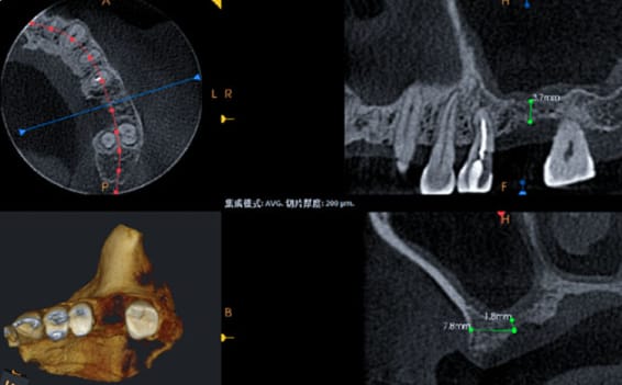
Imaging showed severe vertical deficiency at site 26, with residual ridge height below 4 mm and a buccal bone defect.
CBCT-based planning supported a transcrestal sinus-floor elevation with simultaneous implant placement.
Surgical treatment
Autogenous bone was collected at low speed, followed by osteotome elevation of the sinus floor and ridge expansion.
PRF and autogenous bone particles were placed beneath the elevated sinus membrane. An Excellence Series BL.RC4110 implant (Ø4.1 × 10 mm) was inserted with acceptable initial stability and a healing abutment was fitted.
Restoration and review
Postoperative imaging showed favorable implant position. At five months, the peri-implant condition was good and restorative procedures began.
A BL.MA.RC4625 straight abutment and zirconia all-ceramic crown were used. One year after restoration, the implant remained stable with no reported surrounding abnormality.
This case has been edited and reorganized from the original Trusyou clinical record for clearer professional reading.
View original source