
Patient and presentation
A 34-year-old patient sought treatment for a tooth that had been missing for more than one year and was affecting chewing.
The recorded medical review was unremarkable, with no smoking or alcohol history and no reported systemic contraindications or allergies.
Clinical assessment
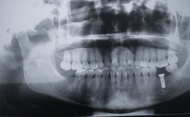
The opposing dentition was normal, and imaging showed no obvious alveolar-bone resorption at site 36.
The diagnosis was a missing mandibular left first molar, and a single implant-supported restoration was planned.
Surgical treatment
A BL.RC4112 implant (Ø4.1 × 12 mm) was placed at site 36 with a BL.HA.RC6020 healing abutment (Ø6.0, H2). The operation used infiltration anesthesia and an I-shaped incision.
The bone was recorded as Class III. No graft material was used, stability was good and immediate postoperative imaging showed favorable three-dimensional positioning with initial stability above 35 Ncm.
Restoration and review
The restorative phase recorded adequate clearance, a stable abutment and close marginal adaptation of the cemented restoration.
Five years after restoration, the implant position and bone level were stable in the available images, with no reported abnormality around the abutment or prosthesis.
This case has been edited and reorganized from the original Trusyou clinical record for clearer professional reading.
View original source