
Patient and presentation
A 57-year-old patient reported a fractured lower-right posterior tooth for three months with impaired chewing.
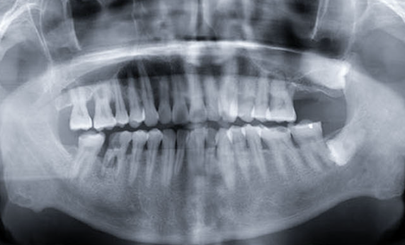
Examination showed a residual crown at 46, extensive caries, mobility, inflamed gingiva and furcation involvement. Imaging showed periapical radiolucency and distal-lingual bone loss.
Site preservation
Tooth 46 was extracted and the socket was debrided. The record describes apical granulomatous tissue and lingual bone perforation.
A-PRF blocks and an A-PRF membrane were used for socket preservation. Early review showed soft-tissue integration around the grafted area.
Delayed implant placement
Before implant placement, bone height and width were considered sufficient despite incomplete remodeling of the former furcation region.
An Excellence Series BL.RC4112 implant (Ø4.1 × 12 mm) with a BL.HA.RC6020 healing abutment was placed at site 46.
Restoration and review
Restoration was completed four months after implantation. Follow-up imaging showed increasing bone density and height around the surgical area.
At 18 months after restoration, the implant was not mobile, surrounding tissues were reported in good condition and the regenerated bone appeared mature and stable.
This case has been edited and reorganized from the original Trusyou clinical record for clearer professional reading.
View original source